Venous Ultrasound
For Appointments Call: 786-332-4106 Miami 954-893-3811 Broward
 Book for $85.00
Venous Ultrasound (Extremities)
- What is Venous Ultrasound Imaging?
- What are some common uses of the procedure?
- How should I prepare?
- What does the equipment look like?
- How does the procedure work?
- How is the procedure performed?
- What will I experience during and after the procedure?
- Who interprets the results and how do I get them?
- What are the benefits vs. risks?
- What are the limitations of Venous Ultrasound Imaging?
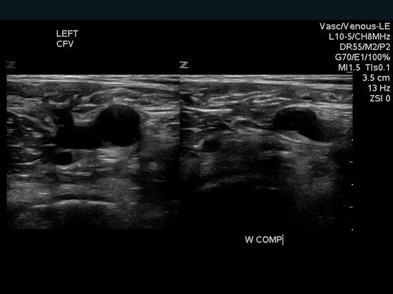
What is Venous Ultrasound Imaging?
Ultrasound imaging, also called ultrasound scanning or sonography, involves the use of a small transducer (probe) and ultrasound gel to expose the body to high-frequency sound waves. Ultrasound is safe and painless, and produces pictures of the inside of the body using sound waves. Ultrasound examinations do not use ionizing radiation (as used in x-rays). Because ultrasound images are captured in real-time, they can show the structure and movement of the body's internal organs, as well as blood flowing through blood vessels.
Ultrasound imaging is a noninvasive medical test that helps physicians diagnose and treat medical conditions.
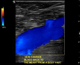
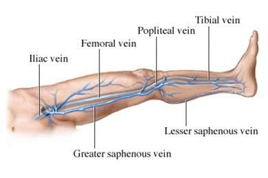
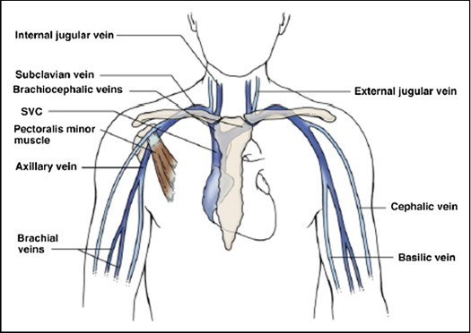
Venous ultrasound provides pictures of the veins throughout the body.
A Doppler ultrasound study may be part of a venous ultrasound examination.
Doppler ultrasound is a special ultrasound technique that evaluates blood flow through a blood vessel, including the body's major arteries and veins in the abdomen, arms, legs and neck.


What are some common uses of the procedure?
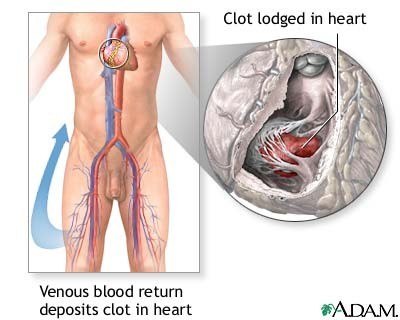
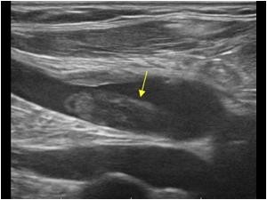
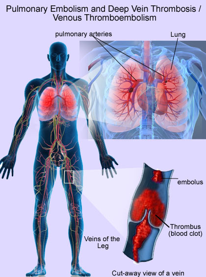
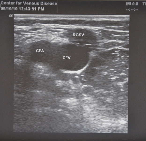
The most common reason for a venous ultrasound exam is to search for blood clots, especially in the veins of the leg. This condition is often referred to as deep vein thrombosis or DVT. These clots may break off and pass into the lungs, where they can cause a dangerous condition called pulmonary embolism. If the blood clot in the leg is found early enough, treatment can be started to prevent it from passing to the lung.
A venous ultrasound study is also performed to:
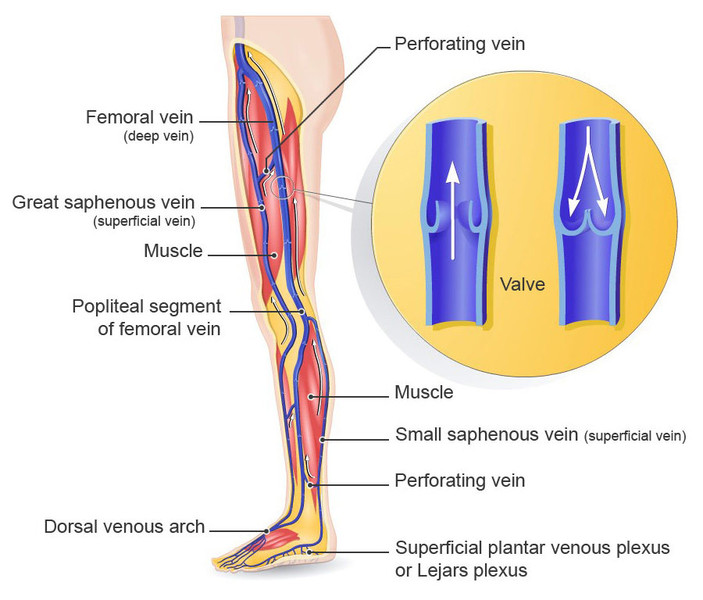
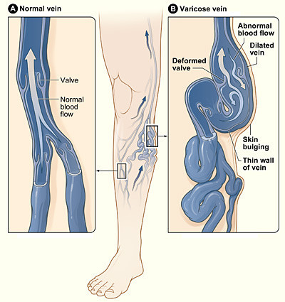
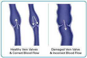
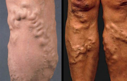
- determine the cause of long-standing leg swelling. In people with a common condition called varicose veins, the valves that keep blood flowing back to the heart in the right direction may be damaged, and venous ultrasound can help the radiologist decide how best to deal with this condition.
- aid in the placement of a needle or catheter into a vein. Sonography can help locate the exact site of the vein and avoid complications, such as bleeding.
- map out the veins in the leg or arm so that pieces of vein may be removed and used to bypass a narrowed or blocked blood vessel. An example is using pieces of vein from the leg to surgically bypass narrowed heart (coronary) arteries.
- examine a blood vessel graft used for dialysis if it is not working as expected; for example, the graft may be narrowed or blocked.
In children, ultrasound is used to:
- aid in the placement of a needle or catheter into a vein to help avoid complications such as bleeding.
- evaluate a connection between an artery and a vein which can be seen in congenital vascular malformations (arteriovenous malformations or fistula) and in dialysis fistula.
If a line is placed in a vein of the legs or arms, there is a much higher chance of developing a clot around it due to the smaller vessel size (especially in infants and young children). In some instances, a clot can form in the arm and extend into the left leg or into the major vein of the abdomen due to compression at the inlet of the chest.
Doppler ultrasound images can help the physician to see and evaluate:
- blockages to blood flow (such as clots).
- narrowing of vessels.
- tumors and congenital vascular malformation.

How should I prepare?
You should wear comfortable, loose-fitting clothing for your ultrasound exam. You may need to remove all clothing and jewelry in the area to be examined.
You may be asked to wear a gown during the procedure.
A period of fasting is necessary only if you are to have an examination of veins in your abdomen. In this case, you will probably be asked not to ingest any food or fluids except water for six to eight hours ahead of time. Otherwise, there is no other special preparation for a venous ultrasound.
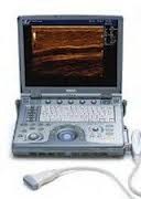
What does the equipment look like?
Ultrasound scanners consist of a console containing a computer and electronics, a video display screen and a transducer that is used to do the scanning. The transducer is a small hand-held device that resembles a microphone, attached to the scanner by a cord. The transducer sends out inaudible high frequency sound waves into the body and then listens for the returning echoes from the tissues in the body. The principles are similar to sonar used by boats and submarines.
The ultrasound image is immediately visible on a video display screen that looks like a computer or television monitor. The image is created based on the amplitude (loudness), frequency (pitch) and time it takes for the ultrasound signal to return from the area of the patient being examined to the transducer, as well as the composition of body tissue through which and the type of body structure the sound travels through.
How does the procedure work?
Ultrasound imaging is based on the same principles involved in the sonar used by bats, ships and fishermen. When a sound wave strikes an object, it bounces back, or echoes. By measuring these echo waves, it is possible to determine how far away the object is and its size, shape and consistency (whether the object is solid or filled with fluid).
In medicine, ultrasound is used to detect changes in appearance of organs, tissues, and vessels or detect abnormal masses, such as tumors.
In an ultrasound examination, a transducer both sends the sound waves and receives the echoing waves. When the transducer is pressed against the skin, it directs small pulses of inaudible, high-frequency sound waves into the body. As the sound waves bounce off of internal organs, fluids and tissues, the sensitive microphone in the transducer records tiny changes in the sound's pitch and direction. These signature waves are instantly measured and displayed by a computer, which in turn creates a real-time picture on the monitor. One or more frames of the moving pictures are typically captured as still images. Small loops of the moving “real time” images may also be saved.
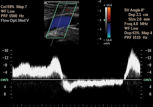
Doppler ultrasound, a special application of ultrasound, measures the direction and speed of blood cells as they move through vessels. The movement of blood cells causes a change in pitch of the reflected sound waves (called the Doppler Effect). A computer collects and processes the sounds and creates graphs or color pictures that represent the flow of blood through the blood vessels.
 Venous Ultrasound
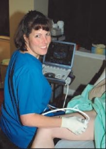
How is the procedure performed?
For most ultrasound exams, the patient is positioned lying face-up on an examination table that can be tilted or moved.
A clear water-based gel is applied to the area of the body being studied to help the transducer make secure contact with the body and eliminate air pockets between the transducer and the skin that can block the sound waves from passing into your body. The sonographer (ultrasound technologist) or radiologist then presses the transducer firmly against the skin in various locations, sweeping over the area of interest or angling the sound beam from a farther location to see an area of concern better.
Doppler sonography is performed using the same transducer.
When the examination is complete, the patient may be asked to dress and wait while the ultrasound images are reviewed.
This ultrasound examination is usually completed within 30 to 45 minutes.
What will I experience during and after the procedure?
Ultrasound examinations are painless, fast and easily tolerated by most patients.
After you are positioned on the examination table, the radiologist or sonographer will apply some warm water-based gel on your skin and then place the transducer firmly against your body, moving it back and forth over the area of interest until the desired images are captured. There is usually no discomfort from pressure as the transducer is pressed against the area being examined.
If scanning is performed over an area of tenderness, you may feel pressure or minor pain from the transducer.
Ultrasound exams in which the transducer is inserted into an opening of the body may produce minimal discomfort.
If a Doppler ultrasound study is performed, you may actually hear pulse-like sounds that change in pitch as the blood flow is monitored and measured.
Once the imaging is complete, the clear ultrasound gel will be wiped off your skin.
After an ultrasound examination, you should be able to resume your normal activities immediately.
Who interprets the results and how do I get them?
A radiologist, a physician specifically trained to supervise and interpret radiology examinations, will analyze the images and send a signed report to your primary care physician, or to the physician or other healthcare provider who referred you for the exam, who will share the results with you. In some cases the radiologist may discuss results with you at the conclusion of your examination.
Follow-up examinations may be necessary, and your doctor will explain the reason why another exam is needed. Sometimes a follow-up exam is done because a suspicious or questionable finding needs clarification with additional views or a special imaging technique. A follow-up examination may be necessary so that any change in a known abnormality can be monitored over time. Follow-up examinations are sometimes the best way to see if treatment is working or if an abnormality is stable over time.
What are the benefits vs. risks?
Benefits
- Most ultrasound scanning is noninvasive (no needles or injections).
- Occasionally, an ultrasound exam may be temporarily uncomfortable, but it is almost never painful.
- Ultrasound is widely available, easy-to-use and less expensive than other imaging methods.
- Ultrasound imaging is extremely safe and does not use any ionizing radiation.
- Ultrasound scanning gives a clear picture of soft tissues that do not show up well on x-ray images.
- Venous ultrasound helps to detect blood clots in the veins of the legs before they become dislodged and pass to the lungs. It can also show the movement of blood within blood vessels.
- Compared to venography, which requires injecting contrast material into a vein, venous ultrasound is accurate for detecting blood clots in the veins of the thigh up to the knee. In the calf, because the veins become very small, ultrasound is less accurate. However, potentially dangerous venous clots are lodged in the larger veins.
Risks
- For standard diagnostic ultrasound, there are no known harmful effects on humans.
What are the limitations of Venous Ultrasound Imaging?
Veins lying deep beneath the skin, especially small veins in the calf, may be hard to see.