
Pelvic Ultrasound
For Appointments Call: 786-332-4106 Miami 954-893-3811 Broward
 Book for $85.00
Ultrasound Imaging of the Pelvis
Pelvic and Obstetric Ultrasound
- What is Ultrasound Imaging of the Pelvis?
- What are some common uses of the procedure?
- How should I prepare?
- What does the equipment look like?
- How does the procedure work?
- How is the procedure performed?
- What will I experience during and after the procedure?
- Who interprets the results and how do I get them?
- What are the benefits vs. risks?
- What are the limitations of Pelvic Ultrasound Imaging?
What is Pelvic Ultrasound Imaging?
Ultrasound imaging, also called ultrasound scanning or sonography, involves the use of a small transducer (probe) and ultrasound gel to expose the body to high-frequency sound waves. Ultrasound is safe and painless, and produces pictures of the inside of the body using sound waves. Ultrasound examinations do not use ionizing radiation (as used in x-rays). Because ultrasound images are captured in real-time, they can show the structure and movement of the body's internal organs, as well as blood flowing through blood vessels.
Ultrasound imaging is a noninvasive medical test that helps physicians diagnose and treat medical conditions.
A pelvic ultrasound provides pictures of the structures and organs in the lower abdomen and pelvis.
There are three types of pelvic ultrasound:
- abdominal (transabdominal)
- vaginal (transvaginal, endovaginal) for women
- rectal (transrectal) for men
A Doppler ultrasound exam may be part of a pelvic ultrasound examination.
Doppler ultrasound is a special ultrasound technique that evaluates blood flow through a blood vessel, including the body's major arteries and veins in the abdomen, arms, legs and neck.
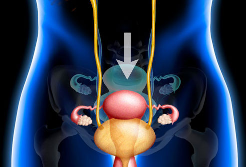
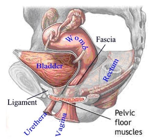
 Female Reproductive System
What are some common uses of the procedure?
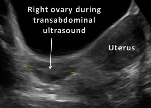
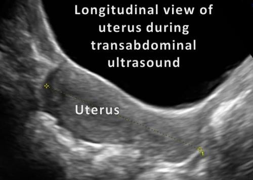
In women, a pelvic ultrasound is most often performed to evaluate the:
- Uterus.
- Cervix.
- Ovaries.
- Fallopian tubes.
- Bladder.
Pelvic ultrasound exams are also used to monitor the health and development of an embryo or fetus during pregnancy. See the Obstetrical Ultrasound page (www.RadiologyInfo.org/en/info.cfm?pg=obstetricus) for more information.


Ultrasound examinations can help diagnose symptoms experienced by women such as:
- pelvic pain
- abnormal bleeding
- other menstrual problems
and help identify:
- palpable masses such as ovarian cysts and uterine fibroids
- ovarian or uterine cancers
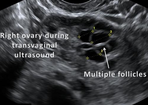
A transvaginal ultrasound is usually performed to view the endometrium or the lining and thickness of the uterine cavity and the ovaries. Transvaginal ultrasound also provides a good way to evaluate the muscular walls of the uterus, called the myometrium. Sonohysterography allows for a more in-depth investigation of the uterine cavity. Three-dimensional (3-D) ultrasound permits evaluation of the uterus and ovaries in planes that cannot be imaged directly. These exams are typically performed to detect:
- uterine anomalies
- uterine scars
- endometrial polyps
- fibroids
- cancer, especially in patients with abnormal uterine bleeding
Some physicians also use 3-D ultrasound or sonohysterography for patients with infertility. Three-dimensional ultrasound provides information about the outer contour of the uterus and about uterine irregularities.
In men, a pelvic ultrasound is used to evaluate the:
- bladder
- seminal vesicles
- prostate
Transrectal ultrasound, a special study usually done to view the prostate gland, involves inserting a specialized ultrasound transducer into a man's rectum. See the Prostate Ultrasound page (www.RadiologyInfo/en/info.cfm?pg=us-prostate) for more information.
In men and women, a pelvic ultrasound exam can help identify:
- kidney stones.
- bladder tumors.
- other disorders of the urinary bladder.
In children, pelvic ultrasound can help evaluate:
- pelvic masses.
- pelvic pain.
- ambiguous genitalia and anomalies of pelvic organs.
- early or delayed puberty in girls.
Pelvic ultrasound is also used to guide procedures such as needle biopsies, in which needles are used to extract a sample of cells from organs for laboratory testing.
Doppler ultrasound images can help the physician to see and evaluate:
- blockages to blood flow (such as clots).
- narrowing of vessels.
- tumors and congenital vascular malformation.
How should I prepare?
You should wear comfortable, loose-fitting clothing for your ultrasound exam. You may need to remove all clothing and jewelry in the area to be examined.
You may be asked to wear a gown during the procedure.
Ultrasound examinations are very sensitive to motion, and an active or crying child will slow the examination process. To ensure a smooth experience, it would be beneficial to explain the procedure to the child prior to the exam. You may bring a book to read to the child to ease anxiety. Ultrasound departments often have a television in the examination room and the child's favorite show may be played if there are no other available distractions.
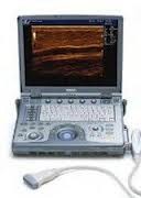
What does the equipment look like?
Ultrasound scanners consist of a console containing a computer and electronics, a video display screen and a transducer that is used to do the scanning. The transducer is a small hand-held device that resembles a microphone, attached to the scanner by a cord. The transducer sends out inaudible high frequency sound waves into the body and then listens for the returning echoes from the tissues in the body. The principles are similar to sonar used by boats and submarines.
The ultrasound image is immediately visible on a video display screen that looks like a computer or television monitor. The image is created based on the amplitude (loudness), frequency (pitch) and time it takes for the ultrasound signal to return from the area of the patient being examined to the transducer, as well as the composition of body tissue through which and the type of body structure the sound travels through.
For ultrasound procedures requiring insertion of the transducer, such as transvaginal or transrectal exams, the device is covered with a sheath and lubricated before insertion.
How does the procedure work?
Ultrasound imaging is based on the same principles involved in the sonar used by bats, ships and fishermen. When a sound wave strikes an object, it bounces back, or echoes. By measuring these echo waves, it is possible to determine how far away the object is and its size, shape and consistency (whether the object is solid or filled with fluid).
In medicine, ultrasound is used to detect changes in appearance of organs, tissues, and vessels or detect abnormal masses, such as tumors.
In an ultrasound examination, a transducer both sends the sound waves and receives the echoing waves. When the transducer is pressed against the skin, it directs small pulses of inaudible, high-frequency sound waves into the body. As the sound waves bounce off of internal organs, fluids and tissues, the sensitive microphone in the transducer records tiny changes in the sound's pitch and direction. These signature waves are instantly measured and displayed by a computer, which in turn creates a real-time picture on the monitor. One or more frames of the moving pictures are typically captured as still images. Small loops of the moving “real time” images may also be saved.
The same principles apply to ultrasound procedures such as transrectal and transvaginal which require insertion of a special transducer into the body.
Doppler ultrasound, a special application of ultrasound, measures the direction and speed of blood cells as they move through vessels. The movement of blood cells causes a change in pitch of the reflected sound waves (called the Doppler Effect). A computer collects and processes the sounds and creates graphs or color pictures that represent the flow of blood through the blood vessels.
How is the procedure performed?
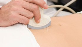
Transabdominal:
For most ultrasound exams, the patient is positioned lying face-up on an examination table that can be tilted or moved.
A clear water-based gel is applied to the area of the body being studied to help the transducer make secure contact with the body and eliminate air pockets between the transducer and the skin that can block the sound waves from passing into your body. The sonographer (ultrasound technologist) or radiologist then presses the transducer firmly against the skin in various locations, sweeping over the area of interest or angling the sound beam from a farther location to see an area of concern better.
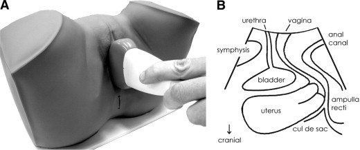
Transvaginal:
Transvaginal ultrasound is performed very much like a gynecologic exam and involves the insertion of the transducer into the vagina after the patient empties her bladder. The tip of the transducer is smaller than the standard speculum used when performing a Pap test. A protective cover is placed over the transducer, lubricated with a small amount of gel, and then inserted into the vagina. Only two to three inches of the transducer end are inserted into the vagina. The images are obtained from different orientations to get the best views of the uterus and ovaries. Transvaginal ultrasound is usually performed with the patient lying on her back, possibly with her feet in stirrups similar to a gynecologic exam.
Transrectal:
For a transrectal ultrasound, a protective cover is placed over the transducer. It is lubricated and then placed into the rectum.
The patient usually lies on his or her side, facing away from the examiner, with the knees and hips slightly flexed.
Doppler sonography is performed using the same transducer.
When the examination is complete, the patient may be asked to dress and wait while the ultrasound images are reviewed.
These ultrasound examinations are usually completed within 30 minutes.
What will I experience during and after the procedure?
Ultrasound examinations are painless, fast and easily tolerated by most patients.
For a transabdominal exam:
After you are positioned on the examination table, the radiologist or sonographer will apply some warm water-based gel on your skin and then place the transducer firmly against your body, moving it back and forth over the area of interest until the desired images are captured. There is usually no discomfort from pressure as the transducer is pressed against the area being examined.
If scanning is performed over an area of tenderness, you may feel pressure or minor pain from the transducer.
For a transvaginal exam:
With transvaginal ultrasound, although the examination is often performed to look for a cause of pelvic pain, the sonogram itself should not be painful or significantly increase your discomfort. A vaginal sonogram is usually more comfortable than a manual gynecologic examination.
For a transrectal exam:
If no biopsy is required, transrectal ultrasound of the prostate is similar or may have less discomfort than a rectal exam performed by your doctor.
If a biopsy is performed, additional discomfort, due to the needle insertion, is usually minimal because the rectal wall is relatively insensitive to the pain in the region of the prostate. A biopsy will add time to the procedure.
If a Doppler ultrasound study is performed, you may actually hear pulse-like sounds that change in pitch as the blood flow is monitored and measured.
Once the imaging is complete, the clear ultrasound gel will be wiped off your skin.
After an ultrasound examination, you should be able to resume your normal activities immediately.
Who interprets the results and how do I get them?
A radiologist, a physician specifically trained to supervise and interpret radiology examinations, will analyze the images and send a signed report to your primary care physician, or to the physician or other healthcare provider who referred you for the exam, who will share the results with you. In some cases the radiologist may discuss results with you at the conclusion of your examination.
Follow-up examinations may be necessary, and your doctor will explain the reason why another exam is needed. Sometimes a follow-up exam is done because a suspicious or questionable finding needs clarification with additional views or a special imaging technique. A follow-up examination may be necessary so that any change in a known abnormality can be monitored over time. Follow-up examinations are sometimes the best way to see if treatment is working or if an abnormality is stable over time.
What are the benefits vs. risks?
Benefits
- Most ultrasound scanning is noninvasive (no needles or injections).
- Occasionally, an ultrasound exam may be temporarily uncomfortable, but it is almost never painful.
- Ultrasound is widely available, easy-to-use and less expensive than other imaging methods.
- Ultrasound imaging is extremely safe and does not use any ionizing radiation.
- Ultrasound scanning gives a clear picture of soft tissues that do not show up well on x-ray images.
- Ultrasound is the preferred imaging modality for the diagnosis and monitoring of pregnant women and their unborn babies.
- Ultrasound provides real-time imaging, making it a good tool for guiding minimally invasive procedures such as needle biopsies and needle aspiration.
- Pelvic ultrasound can help to identify and evaluate a variety of urinary and reproductive system disorders in both sexes without even the minimal risks associated with x-ray exposure.
Risks
- For standard diagnostic ultrasound, there are no known harmful effects on humans.
What are the limitations of Pelvic Ultrasound Imaging?
Ultrasound waves are disrupted by air or gas; therefore ultrasound is not an ideal imaging technique for air-filled bowel or organs obscured by the bowel. In most cases, barium exams, CT scanning, and MRI are the methods of choice in this setting.
Large patients are more difficult to image by ultrasound because greater amounts of tissue attenuates (weakens) the sound waves as they pass deeper into the body.