
Carotid Ultrasound Imaging
For Appointments Call: 786-332-4106 Miami 954-893-3811 Broward
 Book for $85.00
Carotid Ultrasound Imaging
- What is Ultrasound Imaging of the Carotid Artery?
- What are some common uses of the procedure?
- How should I prepare?
- What does the equipment look like?
- How does the procedure work?
- How is the procedure performed?
- What will I experience during and after the procedure?
- Who interprets the results and how do I get them?
- What are the benefits vs. risks?
- What are the limitations of Carotid Ultrasound Imaging?
What is Carotid Ultrasound Imaging?
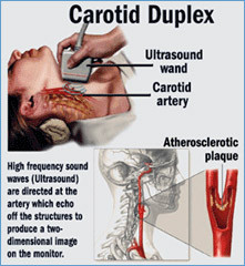
Ultrasound imaging, also called ultrasound scanning or sonography, involves the use of a small transducer (probe) and ultrasound gel to expose the body to high-frequency sound waves. Ultrasound is safe and painless, and produces pictures of the inside of the body using sound waves. Ultrasound examinations do not use ionizing radiation (as used in x-rays). Because ultrasound images are captured in real-time, they can show the structure and movement of the body's internal organs, as well as blood flowing through blood vessels.
Ultrasound imaging is a noninvasive medical test that helps physicians diagnose and treat medical conditions.
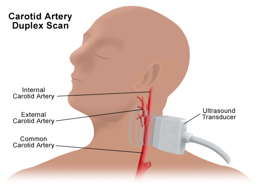
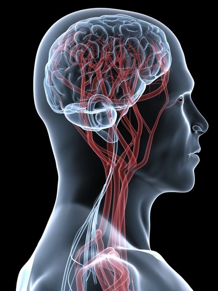
An ultrasound of the body's two carotid arteries, which are located on each side of the neck and carry blood from the heart to the brain, provides detailed pictures of these blood vessels and information about the blood flowing through them.
A Doppler ultrasound study is usually an integral part of a carotid ultrasound examination.
Doppler ultrasound is a special ultrasound technique that evaluates blood flow through a blood vessel, including the body's major arteries and veins in the abdomen, arms, legs and neck.


What are some common uses of the procedure?
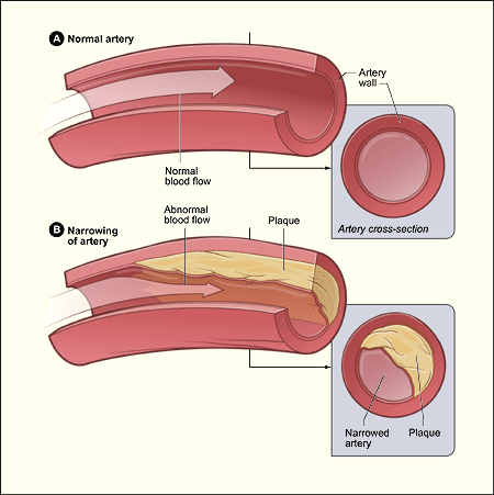
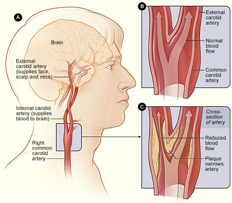
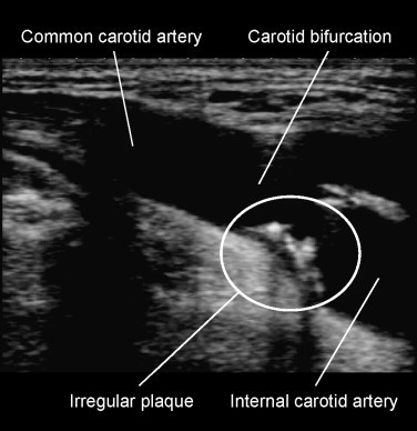
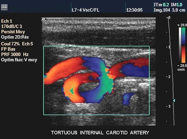
The carotid ultrasound is most frequently performed to detect narrowing, or stenosis, of the carotid artery, a condition that substantially increases the risk of stroke.
The major goal of carotid ultrasound is to screen patients for blockage or narrowing of their carotid arteries, which if present may increase their risk of having a stroke. If a significant narrowing is detected, a comprehensive treatment may be initiated.
It may also be performed if a patient has high blood pressure or a carotid bruit (pronounced brU-E)—an abnormal sound in the neck that is heard with the stethoscope. In some cases, it is also performed in preparation for a coronary artery bypass. Other risk factors calling for a carotid ultrasound are:
- diabetes
- elevated blood cholesterol
- a family history of stroke or heart disease
A carotid ultrasound is also performed to:
- locate a hematoma, a collection of clotted blood that may slow and eventually stop blood flow.
- check the state of the carotid artery after surgery to restore normal blood flow.
- verify the position of a metal stent placed to maintain carotid blood flow.
Doppler ultrasound images can help the physician to see and evaluate:
- blockages to blood flow (such as clots).
- narrowing of vessels.
- tumors and congenital vascular malformation.
In children, Doppler ultrasound is used to:
- evaluate blood flow.
- predict a higher risk of stroke in children with sickle cell disease.
detect abnormalities in the blood vessels, lymph nodes and lymphatic vessels.
How should I prepare?
You should wear comfortable, loose-fitting clothing for your ultrasound exam. You may need to remove all clothing and jewelry in the area to be examined.
A loose-fitting, open necked shirt or blouse is ideal.
Ultrasound examinations are very sensitive to motion, and an active or crying child will slow the examination process. To ensure a smooth experience, it would be beneficial to explain the procedure to the child prior to the exam. You may bring a book to read to the child to ease anxiety. Ultrasound departments often have a television in the examination room and the child's favorite show may be played if there are no other available distractions.
No other preparation is required.
What does the equipment look like?
Ultrasound scanners consist of a console containing a computer and electronics, a video display screen and a transducer that is used to do the scanning. The transducer is a small hand-held device that resembles a microphone, attached to the scanner by a cord. The transducer sends out inaudible high frequency sound waves into the body and then listens for the returning echoes from the tissues in the body. The principles are similar to sonar used by boats and submarines.
The ultrasound image is immediately visible on a video display screen that looks like a computer or television monitor. The image is created based on the amplitude (loudness), frequency (pitch) and time it takes for the ultrasound signal to return from the area of the patient being examined to the transducer, as well as the composition of body tissue through which and the type of body structure the sound travels through.
How does the procedure work?
Ultrasound imaging is based on the same principles involved in the sonar used by bats, ships and fishermen. When a sound wave strikes an object, it bounces back, or echoes. By measuring these echo waves, it is possible to determine how far away the object is and its size, shape and consistency (whether the object is solid or filled with fluid).
In medicine, ultrasound is used to detect changes in appearance of organs, tissues, and vessels or detect abnormal masses, such as tumors.
In an ultrasound examination, a transducer both sends the sound waves and receives the echoing waves. When the transducer is pressed against the skin, it directs small pulses of inaudible, high-frequency sound waves into the body. As the sound waves bounce off of internal organs, fluids and tissues, the sensitive microphone in the transducer records tiny changes in the sound's pitch and direction. These signature waves are instantly measured and displayed by a computer, which in turn creates a real-time picture on the monitor. One or more frames of the moving pictures are typically captured as still images. Small loops of the moving “real time” images may also be saved
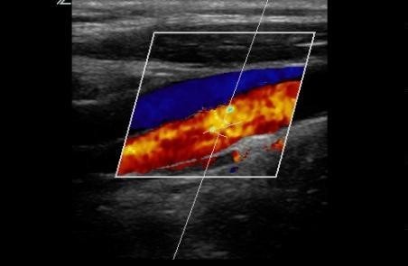
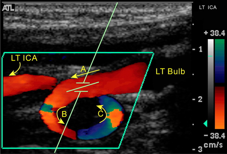
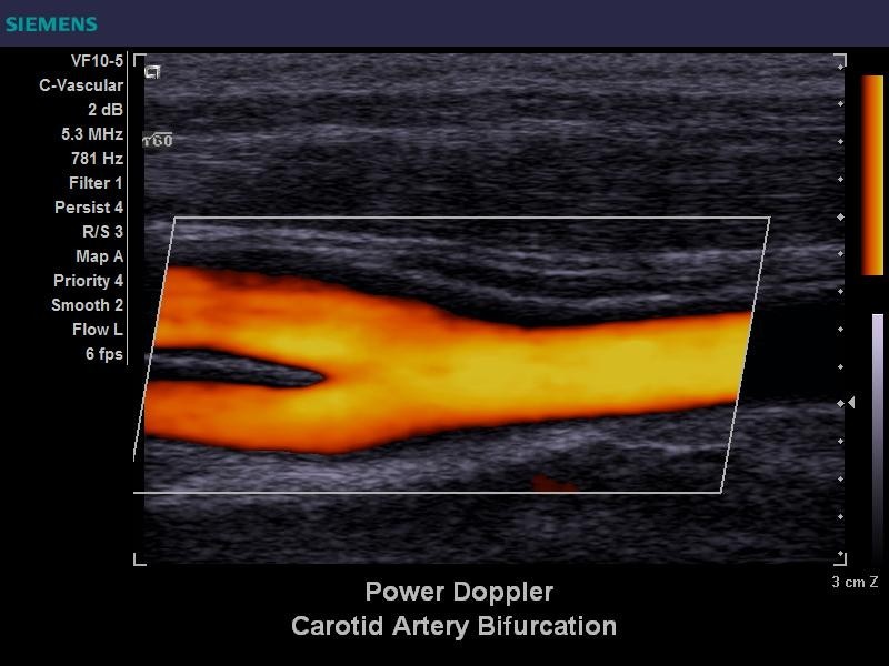
Doppler ultrasound, a special application of ultrasound, measures the direction and speed of blood cells as they move through vessels. The movement of blood cells causes a change in pitch of the reflected sound waves (called the Doppler Effect). A computer collects and processes the sounds and creates graphs or color pictures that represent the flow of blood through the blood vessels.
How is the procedure performed?
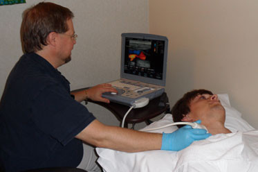
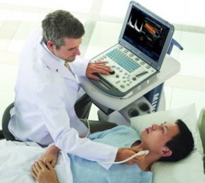
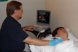
For most ultrasound exams, the patient is positioned lying face-up on an examination table that can be tilted or moved.
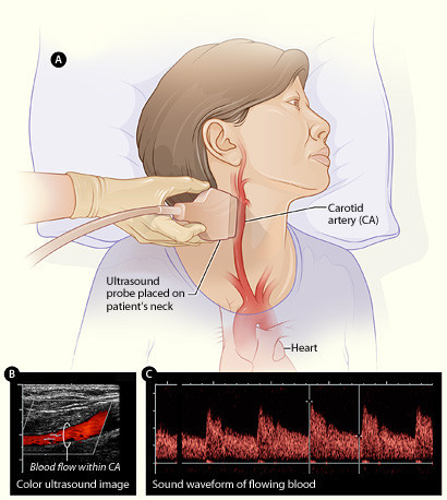
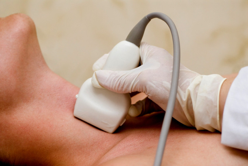
A clear water-based gel is applied to the area of the body being studied to help the transducer make secure contact with the body and eliminate air pockets between the transducer and the skin that can block the sound waves from passing into your body. The sonographer (ultrasound technologist) or radiologist then presses the transducer firmly against the skin in various locations, sweeping over the area of interest or angling the sound beam from a farther location to see an area of concern better.
Doppler sonography is performed using the same transducer.
When the examination is complete, the patient may be asked to dress and wait while the ultrasound images are reviewed.
The branches of the carotid arteries inside the head cannot be seen directly but can be evaluated with Doppler ultrasound in children with sickle cell disease. It is performed by placing the transducer over the child's temple and recording the blood flow in the center of the skull.
This ultrasound examination is usually completed within 30 to 45 minutes.
What will I experience during and after the procedure?
Ultrasound examinations are painless, fast and easily tolerated by most patients.
After you are positioned on the examination table, the radiologist or sonographer will apply some warm water-based gel on your skin and then place the transducer firmly against your body, moving it back and forth over the area of interest until the desired images are captured. There is usually no discomfort from pressure as the transducer is pressed against the area being examined.
If scanning is performed over an area of tenderness, you may feel pressure or minor pain from the transducer.
It may be necessary to tilt or rotate your head for the best exposure, as the transducer is swept over the entire length of your neck on both sides to obtain views of the artery from different perspectives. It also helps to keep your arm and shoulder down. Your head will be supported to keep it still.
If a Doppler ultrasound study is performed, you may actually hear pulse-like sounds that change in pitch as the blood flow is monitored and measured.
Once the imaging is complete, the clear ultrasound gel will be wiped off your skin.
After an ultrasound examination, you should be able to resume your normal activities immediately.
Who interprets the results and how do I get them?
A radiologist, a physician specifically trained to supervise and interpret radiology examinations, will analyze the images and send a signed report to your primary care physician, or to the physician or other healthcare provider who referred you for the exam, who will share the results with you. In some cases the radiologist may discuss results with you at the conclusion of your examination.
Follow-up examinations may be necessary, and your doctor will explain the reason why another exam is needed. Sometimes a follow-up exam is done because a suspicious or questionable finding needs clarification with additional views or a special imaging technique. A follow-up examination may be necessary so that any change in a known abnormality can be monitored over time. Follow-up examinations are sometimes the best way to see if treatment is working or if an abnormality is stable over time.
What are the benefits vs. risks?
Benefits
- Most ultrasound scanning is noninvasive (no needles or injections).
- Occasionally, an ultrasound exam may be temporarily uncomfortable, but it is almost never painful.
- Ultrasound is widely available, easy-to-use and less expensive than other imaging methods.
- Ultrasound imaging is extremely safe and does not use any ionizing radiation.
- Ultrasound scanning gives a clear picture of soft tissues that do not show up well on x-ray images.
- If a carotid ultrasound exam shows narrowing of one or both carotid arteries, treatment can be taken to restore the free flow of blood to the brain. Many strokes are prevented as a result.
Risks
- For standard diagnostic ultrasound, there are no known harmful effects on humans.
- In nearly 50 years of experience, carotid ultrasound has proved to be a risk-free procedure.
What are the limitations of Carotid Ultrasound Imaging?
- Carotid ultrasound may be difficult or impossible if a patient has a dressing covering a wound or surgical scar in the neck.
- An occasional patient is difficult to examine because of the size or contour of the neck.
- Calcium deposits in the wall of the carotid artery may make it difficult to evaluate the vessel.
- A small amount of soft plaque that produces low-level echoes may go undetected.
Ultrasound cannot visualize the entire length of the vessel because the last portion of the carotid artery travels though the bone at the base of the skull. For a more complete assessment, patients may need to undergo a CT or MRI of the carotid arteries.