What is Spinal X-Ray?
For Appointments Call: 786-332-4106 Miami 954-893-3811 Broward
 Book for $70.00
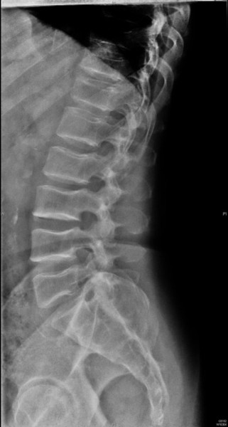
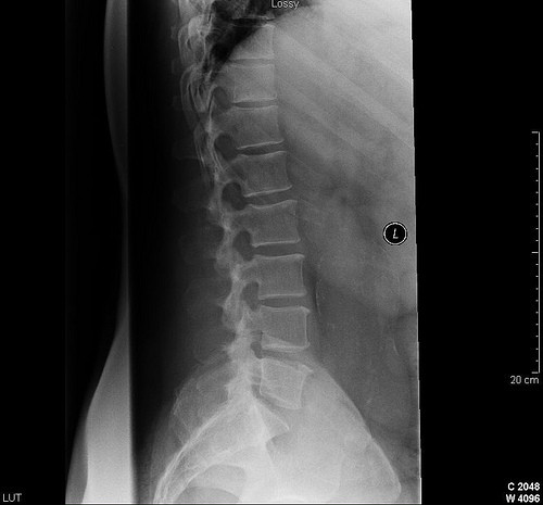
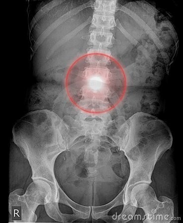
Spinal X-Ray
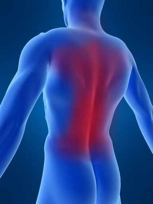
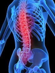
Spinal X-rays are pictures of the spine. They may be taken to find injuries or diseases that affect the discs or joints in your spine. These problems may include spinal fractures, infections, dislocations, tumors, bone spurs, or disc disease.
Spinal X-rays are also done to check the curve of your spine (scoliosis) or for spinal defects.
X-rays are a form of radiation, like light or radio waves, that are focused into a beam, much like a flashlight beam. X-rays can pass through most objects, including the human body.
Dense tissues in the body, such as bones, block (absorb) many of the X-rays and look white on an X-ray picture. Less dense tissues, such as muscles and organs, block fewer of the X-rays (more of the X-rays pass through) and look like shades of gray on an X-ray. X-rays that pass only through air look black on the picture.
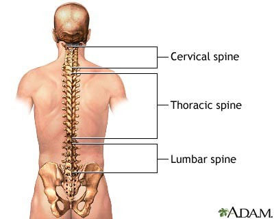
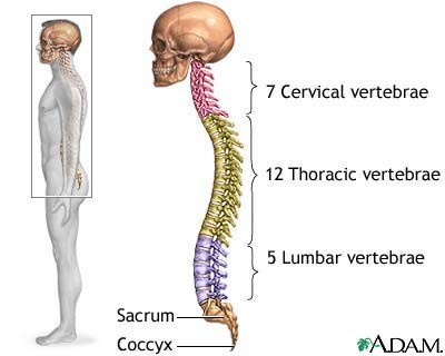
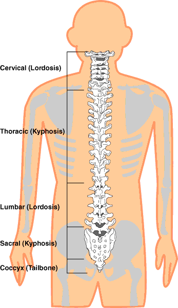
The spine is divided into four parts. So there are four common types of spinal X-rays:
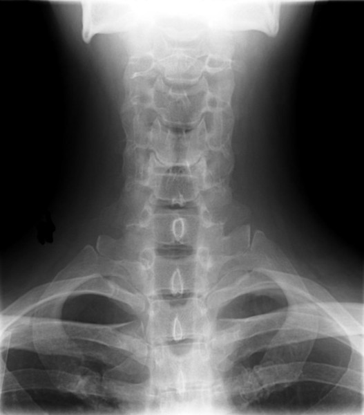
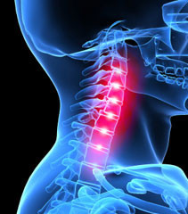
- Cervical spine X-ray. This X-ray test takes pictures of the 7 neck (cervical) bones.
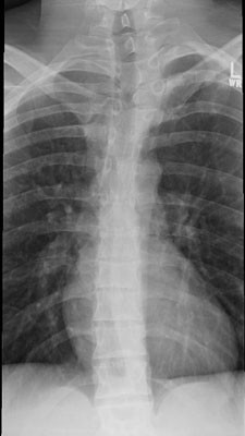
- Thoracic spine X-ray. This X-ray test takes pictures of the 12 chest (thoracic) bones.
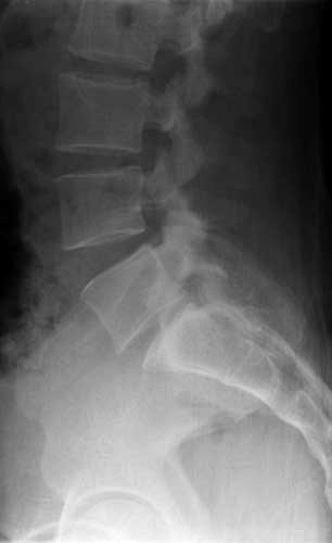
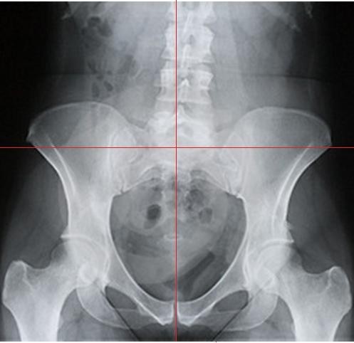
- Lumbosacral spine X-ray. This X-ray test takes pictures of the 5 bones of the lower back (lumbar vertebrae) and a view of the 5 fused bones at the bottom of the spine (sacrum).
- Sacrum/coccyx X-ray. This X-ray test takes a detailed view of the 5 fused bones at the bottom of the spine (sacrum) and the 4 small bones of the tailbone (coccyx).
The most common spinal X-rays are of the cervical vertebrae (C-spine films) and lumbosacral vertebrae (LS-spine films).


Why It Is Done
A spinal X-ray is done to:
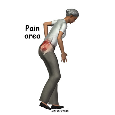
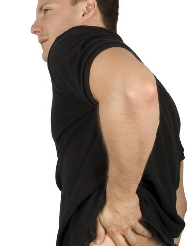
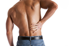
- Find the cause of ongoing pain, numbness, or weakness.
- Check for arthritis of the joints between the vertebrae and the breakdown (degeneration) of the discs between the spinal bones.
- Check injuries to the spine, such as fractures or dislocations.
- Check the spine for effects from other problems, such as infections, tumors, or bone spurs.
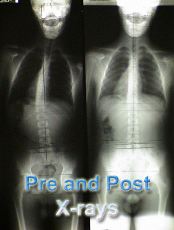
- Check for abnormal curves of the spine, such as scoliosis, in children or young adults.
- Check the spine for problems present at birth (congenital conditions), such as spina bifida, in infants, children, or young adults.
- Check changes in the spine after spinal surgery.
How To Prepare
Before the X-ray test, tell your doctor if you:
- Are or might be pregnant. The risk of radiation exposure to your unborn baby (fetus) must be considered. The risk of damage from the X-rays is usually very low compared with the potential benefits of the test. If a spinal X-ray is absolutely necessary, a lead apron will be placed over your belly to shield your baby from the X-rays.
- Have had an X-ray test using barium contrast material (such as a barium enema) in the past 4 days. Barium shows up on X-ray films and makes it hard to get a clear picture of the lower back (lumbar spine).
You don't need to do anything else before you have this test.
How It Is Done
A spinal X-ray is taken by a radiology technologist. The X-ray pictures are usually read by a doctor who specializes in reading X-rays (radiologist).
You will need to remove any jewelry that may be in the way of the X-ray picture. You may need to take off some of your clothes, depending on which area is examined. You will be given a cloth or paper gown to use during the test. You may be allowed to keep on your underwear if it does not get in the way of the test.
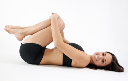
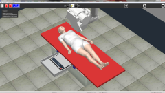
During the X-ray test, you will lie on an X-ray table. If the X-ray is being taken because of a possibly serious injury to your neck or back, to prevent causing more injury a radiologist will look at the first X-ray pictures before taking others.
Usually 3 to 5 X-ray pictures are taken. You need to lie very still to avoid blurring the pictures.
A spinal X-ray usually takes about 15 minutes. You will wait about 5 minutes until the X-rays are processed in case more pictures need to be taken.
How It Feels
You will feel no discomfort from the X-rays. The X-ray table may feel hard, and the room may be cool. You may find that the positions you need to hold are uncomfortable or painful, especially if you have an injury.
Risks
There is always a slight risk of damage to cells or tissue from being exposed to any radiation, including the low levels of radiation used for this test. But the risk of damage from the X-rays is usually very low compared with the potential benefits of the test.
For example, the radiation exposure from a chest X-ray is about equal to the natural radiation exposure received during a round-trip airline flight from Boston to Los Angeles (or Montreal to Vancouver) or 10 days in the Rocky Mountains (Denver, Colorado)
What Affects the Test
Reasons you may not be able to have the test or why the results may not be helpful include:
- If you are pregnant. The X-rays may not be safe for the fetus.
- If you have had a test with barium contrast material in the past 4 days. Barium shows up on X-ray films and can make it hard to get a clear picture.
- If you can't remain still during the test. The pictures may not be clear.
- If you are very overweight. This can make it hard to see the details of the spinal X-ray.

What To Expect After a Spinal X- Ray
- You usually can go back to your normal routine right after a chest x ray.
- A radiologist will analyze, or "read," your x-ray images. This doctor is specially trained to supervise x-ray tests and look at the x-ray pictures.
The radiologist will send a report to your doctor (who requested the x-ray test). Your doctor will discuss the results with you.