
What is a Hip X-Ray?
For Appointments Call: 786-332-4106 Miami 954-893-3811 Broward
 Book for $70.00
What It Is
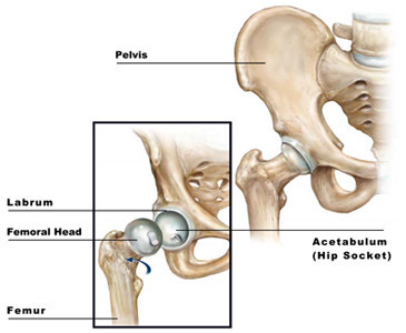
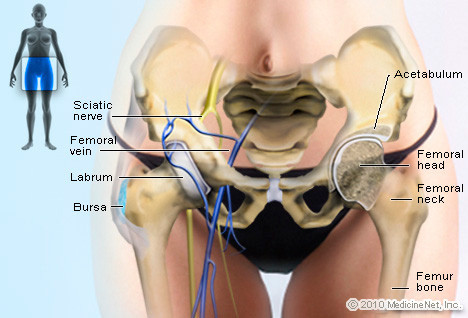
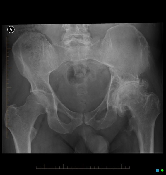
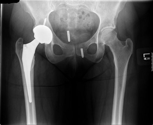
A hip X-ray is a safe and painless test that uses a small amount of radiation to make images of a person's hip joints (where the legs attach to the pelvis). During the examination, an X-ray machine sends a beam of radiation through the pelvic bones and hip joints, and an image is recorded on a computer or special film. This image shows the soft tissues and the bones of the pelvis and hip joints.
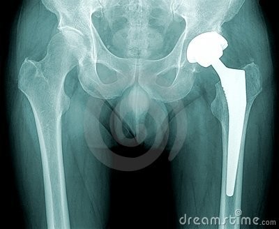
The X-ray image is black and white. Dense body parts that block the passage of the X-ray beam through the body, such as bones, appear white on the X-ray image. Softer body tissues, such as the skin and muscles, allow the X-ray beams to pass through them and appear darker. An X-ray technician takes the X-rays.
An X-ray technician in the radiology department takes the X-rays. Two different pictures are usually taken of the hip: one from the front (anteroposterior view or AP), and one from the side (lateral view, also known as the frog leg lateral view). Typically X-rays of both hips are taken for comparison, even if only one hip is causing the symptoms.


Why It's Done
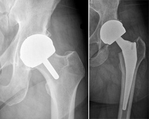
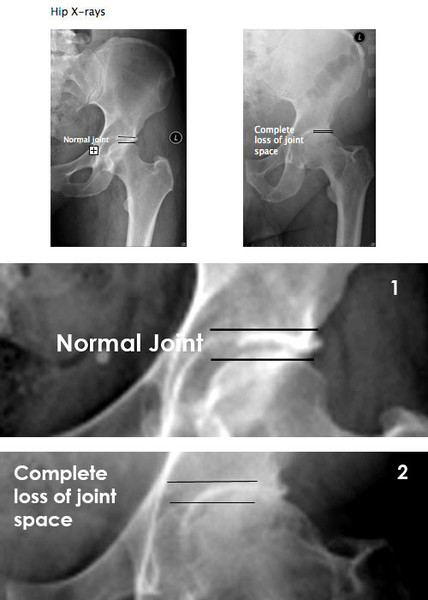
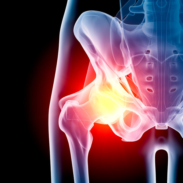
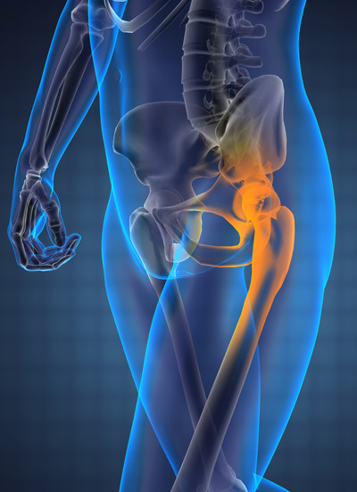
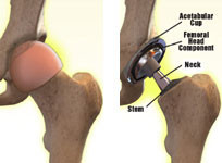
A hip X-ray can help find the cause of common signs and symptoms such as limping, pain, tenderness, swelling, or deformity in the hip area. It can detect broken bones or a dislocated joint. If hip surgery is required, an X-ray may be taken to plan for the surgery and assess the results of the operation.
Also, a hip X-ray can help to detect bone cysts, tumors, infection of the hip joint, or other diseases in the bones of the hips.
Preparation
A hip X-ray doesn't require any special preparation. You may be asked to remove some clothing, jewelry, or any metal objects that might interfere with the image.
If you’re pregnant, it's important to tell the X-ray technician and your doctor. X-rays are usually avoided during pregnancy because there's a small chance the radiation may harm the developing baby. But if the X-ray is necessary, precautions can be taken to protect the fetus.

Procedure
Although the procedure may take about 10 minutes or longer, actual exposure to radiation is usually less than a second.
You may be asked to lie down for this exam. Parents are usually able to accompany their child to provide reassurance. If you stay in the room while the X-ray is being done, you'll be asked to wear a lead apron to protect certain parts of your body. Your child's reproductive organs will also be protected with a lead shield.
The technician or radiologist will position the patient on the table, then step behind a wall or into an adjoining room to operate the machine. Two X-rays are usually taken, one with the legs straight (AP view) and one with the knees apart and feet together (frog leg view), which is how the lateral view usually done. The technician will return to reposition the patient for each X-ray
What to Expect
You won't feel anything as the X-rays are taken. The X-ray room may feel cool due to air conditioning used to maintain the equipment.
The positions required for the X-rays may feel uncomfortable, but they need to be held for only a few seconds. If you have an injury or are in pain and can't stay in the required position, the technician might be able to find another position that's more comfortable.
After the X-rays are taken, you will be asked to wait a few minutes while the images are processed. If they are blurred or unclear, the X-rays may need to be redone.

Getting the Results
The X-rays will be looked at by a radiologist (a doctor who's specially trained in reading and interpreting X-ray images). The radiologist will send a report to your doctor, who will discuss the results with you and explain what they mean. In an emergency, the results of an X-ray can be available quickly. Otherwise, results are usually ready in 24hrs or more.