What is an X-Ray of the Extremitities?
For Appointments Call: 786-332-4106 Miami 954-893-3811 Broward
 Book for $70.00
Extremity X-Ray
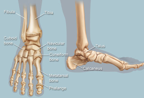
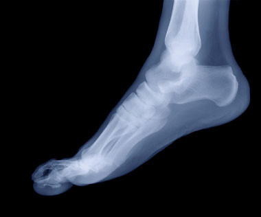
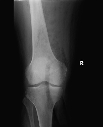
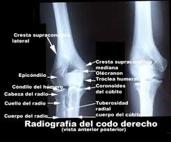
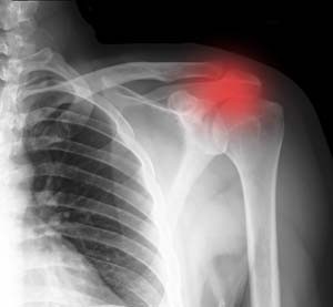
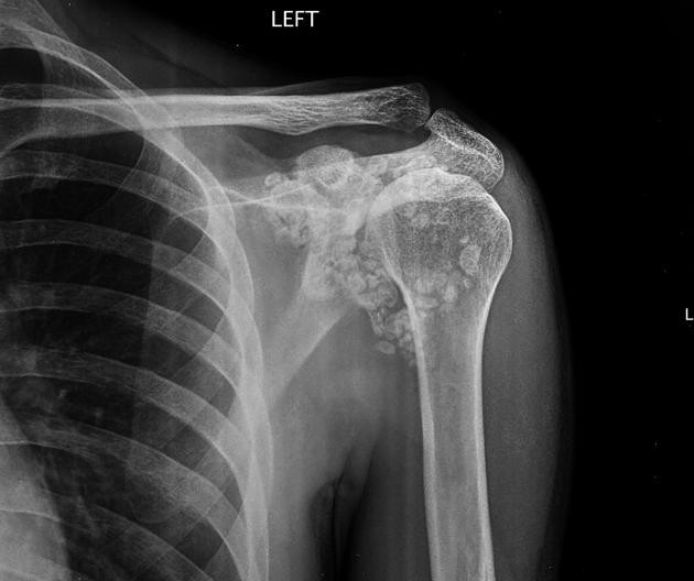
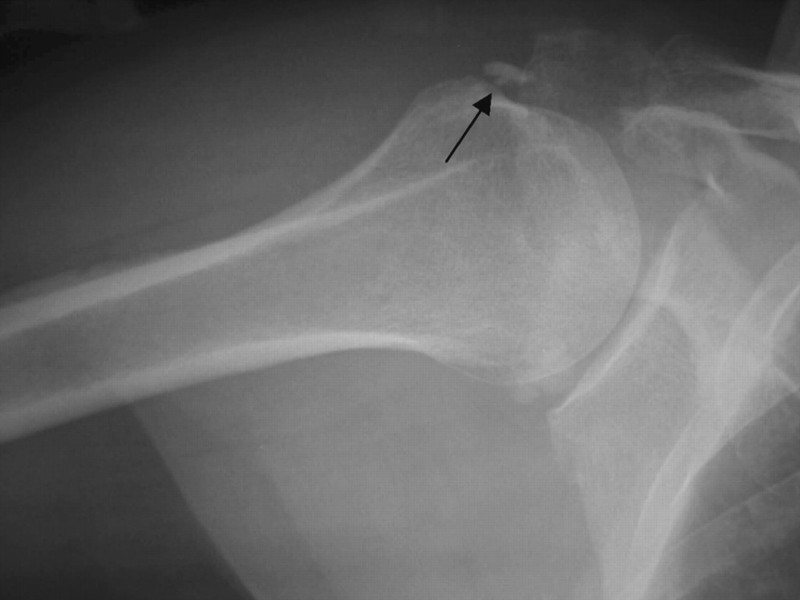
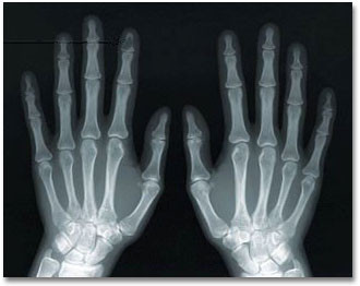
An extremity X-ray is a picture of your hand, wrist, arm, foot, ankle, knee, hip, or leg. It is done to see whether a bone has been fractured or a joint dislocated. It is also used to check for an injury or damage from conditions such as an infection, arthritis, bone growths (tumors), or other bone diseases, such as osteoporosis.
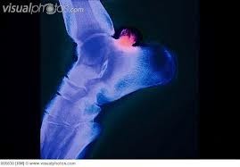
X-rays are a form of radiation, like light or radio waves, that are focused into a beam, much like a flashlight beam. X-rays can pass through most objects, including the human body. X-rays make a picture by striking a detector that either exposes a film or sends the picture to a computer. Dense tissues in the body, such as bones, block (absorb) many of the X-rays and look white on an X-ray picture. Less dense tissues, such as muscles and organs, block fewer of the X-rays (more of the X-rays pass through) and look like shades of gray on an X-ray. X-rays that pass only through air look black on the picture.


Why It Is Done
Extremity X-rays are done to:
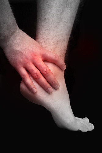
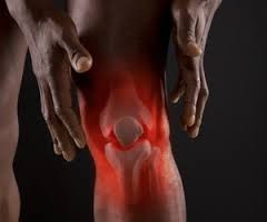
- Find the cause of pain in an extremity.
- See if your bone is fractured or your joint is dislocated.
- See if fluid has built up in the joint or around a bone.
- See if your bones are positioned properly after treatment for a fracture or dislocation, such as after placing a cast or splint on an arm or leg. An X-ray also may be done after a doctor places a device such as a pin or an artificial joint in a bone.
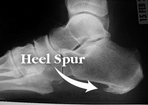
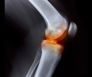
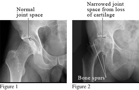
- Find changes in your bones caused by conditions such as an infection, arthritis, bone growths (tumors), or other bone diseases. See pictures of osteoarthritis of the hip and osteoarthritis of the knee .
- Find foreign objects such as pieces of glass or metal.
- Check to see if a child's bones are growing normally.
- See if your bones and joints are in the correct position after joint replacement surgery.
How To Prepare
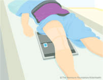
Before the X-ray test, tell your doctor if you are or might be pregnant. Pregnancy and the risk of radiation exposure to your unborn baby (fetus) must be considered. The risk of damage from the X-rays is usually very low compared with the potential benefits of the test. If an extremity X-ray is absolutely necessary, a lead apron will be placed over your abdomen to help shield your baby from exposure to the X-rays.
You don't need to do anything else before you have this test.
How It Is Done
An extremity X-ray is taken by a radiology technologist. The X-ray pictures are usually read by a doctor who specializes in interpreting X-rays (radiologist). Some other types of doctors can also review extremity X-ray pictures for common problems, such as fractures or arthritis.
You will need to remove any jewelry that may be in the way of the X-ray picture. You may need to take off some of your clothes, depending on which area is examined. You will be given a cloth or paper gown to use during the test. You may be allowed to keep on your underwear if it does not get in the way of the test.
During the X-ray test, you will sit by or be on an X-ray table with a film holder under the affected limb. The X-ray technologist will position your limb. If you have an injury, your leg or arm will be handled gently and supported when moved or repositioned. Pillows, sandbags, or other objects may be used to hold the injured limb in place while the pictures are taken. If you are wearing a brace or other device, it may need to be removed. A lead shield may be placed over your pelvic area to protect it from radiation.
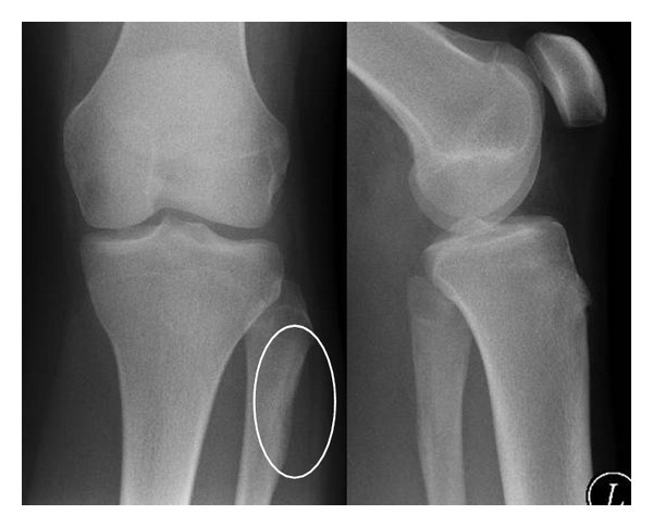
Two or more pictures of the affected limb are usually taken. The test focuses on the specific area that is injured or damaged.
X-ray pictures may also be taken of joints or limbs other than those where the obvious injury has occurred, since an injury at one point may cause damage somewhere else. For example, X-rays of the thighbone (femur) may include pictures of both the knee and hip joints.
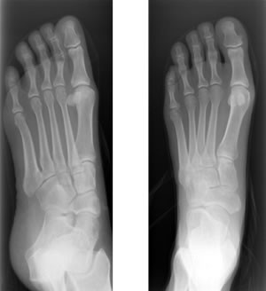
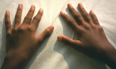
Sometimes an X-ray picture of the unaffected limb is taken so it can be compared with the affected limb. This may happen with children because their bones are still growing. In children, there is an area called a growth plate where new bone is forming. Because it can be difficult to see fractures or other changes in the growth plate, it is helpful to compare the affected limb to the unaffected limb.
An extremity X-ray usually takes about 5 to 10 minutes. You will wait about 5 minutes until the X-rays are processed in case repeat pictures need to be taken
How It Feels
You will feel no discomfort from the X-rays. The X-ray table may feel hard and the room may be cool. You may find that the positions you need to hold are uncomfortable or painful, especially if you have an injury.
Risks
There is always a slight risk of damage to cells or tissue from being exposed to any radiation, including the low levels of radiation used in this test. But the risk of damage from the X-rays is usually very low compared with the potential benefits of the test.
For example, the radiation exposure from a chest X-ray is about equal to the natural radiation exposure received during a round-trip airline flight from Boston to Los Angeles (Montreal to Vancouver) or ten days in the Rocky Mountains (Denver, Colorado).
What Affects the Test
Reasons you may not be able to have the test or why the results may not be helpful include:
- If you can't remain still during the test. This may cause the pictures to be blurry.
- If you are very overweight. This can make it hard to see details in some types of X-ray pictures.
- If you are pregnant and need an X-ray of a leg in the area close to the pelvis.

What To Expect After an extremity Ray
You usually can go back to your normal routine right after the x ray.
A radiologist will analyze, or "read," your x-ray images. This doctor is specially trained to supervise x-ray tests and look at the x-ray pictures.
The radiologist will send a report to your doctor (who requested the x-ray test). Your doctor will discuss the results with you.
In an emergency, you'll get the x-ray results right away. Otherwise, it may take 24 hours or more. Talk with your doctor about when you should expect the results.