
Lower & Upper Extremity Venous
Ultrasound imaging is a noninvasive medical test that helps physicians diagnose and treat medical conditions.
Venous ultrasound provides pictures of the veins throughout the body.
A Doppler ultrasound study may be part of a venous ultrasound examination.
Doppler ultrasound is a special ultrasound technique that evaluates blood flow through a blood vessel, including the body's major arteries and veins in the abdomen, arms, legs and neck.
What are some common uses of the procedure?
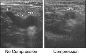
The most common reason for a venous ultrasound exam is to search for blood clots, especially in the veins of the leg. This condition is often referred to as deep vein thrombosis or DVT. These clots may break off and pass into the lungs, where they can cause a dangerous condition called pulmonary embolism. If the blood clot in the leg is found early enough, treatment can be started to prevent it from passing to the lung.
A venous ultrasound study is also performed to:
- determine the cause of long-standing leg swelling. In people with a common condition called varicose veins, the valves that keep blood flowing back to the heart in the right direction may be damaged, and venous ultrasound can help the radiologist decide how best to deal with this condition.
- aid in the placement of a needle or catheter into a vein. Sonography can help locate the exact site of the vein and avoid complications, such as bleeding.
- map out the veins in the leg or arm so that pieces of vein may be removed and used to bypass a narrowed or blocked blood vessel. An example is using pieces of vein from the leg to surgically bypass narrowed heart (coronary) arteries.
- examine a blood vessel graft used for dialysis if it is not working as expected; for example, the graft may be narrowed or blocked.